
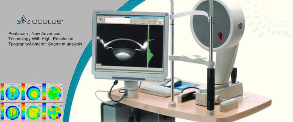
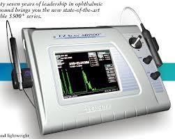
A combination of high frequency, low noise probes and proprietary algorithms provides high-quality B-scan images and precise A-scan measurements of corneal thickness, ACD, lens thickness, and axial length.
A-Scan Probes: 10 MHz, focused, internal fixation light; Solid Tip or Soft-Touch Measurements: ACD, Lens, Vitreous, and Axial Length using individual zone velocities and moving gates Formulas: Holladay, Regression-II, Theoretic/ T, Binkhorst, Hoffer-Q, Haigis (optional)
B-Scan Probes: 10MHz, focused transducer, 30 frames/sec. Measurements: Distance and area Amplifier: 100 dB Gain, Logarithmic/ Linear/S-Curve, Gain, and TVG controls Magnification: Continuous Zoom (0.5x – 2.0x) with Pan (joystick controlled) Display Resolution: 640 x 480 pixels, color VGA with optimal tissue resolution of 0.15mm Processing: Reject below level, enhance contour and texture Freeze: Foot Pedal or touch screen activated Image: B-Scan with simultaneous selectable vector A-Scan Display: 60° sector fan, 128 lines, Gray Scale, B/a presentation (B emphasized) or A/B (A emphasized), Gain TVG, Electronic Scale, Amplifier, OD/OS, Velocity, Probe Orientation, Patient and User Names, Date/Time..
For more information CLICK HERE